General Considerations
The term paraprotein was introduced by Apitz in 1940 and is given to describe the abnormal proteins that are produced by myeloma cells and occur in the blood, urine and tissues.
Paraproteins are the earliest described tumour markers and remain an essential part of the investigation, diagnosis and monitoring of patients with B cell dyscrasias.
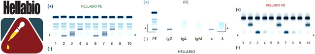
Electrophoresis is the only reliable way of detecting a paraprotein in biological fluids. This test is the initial screening procedure and therefore should have sufficient resolution to do this adequately. Serum electrophoresis should always be accompanied by measurement of serum IgG, IgA and IgM concentrations. Samples with raised IgA and IgM concentrations that cannot be confirmed as polyclonal by the electrophoresis pattern should be analyzed by immunofixation to exclude small paraprotein bands obscured by one of the normal zones.
The majority of serum paraproteins will be found in the region from the start of the beta to the end of the gamma zones. Occasionally paraprotein bands appear in the alpha-zones and in the post gamma region.
Presence of monoclonal free light chains or Bence Jones protein in urine is suggestive of B cell malignancy and may be the only tumour marker. Whatever analytical system is being used, a trace of albumin should be visible in every urine sample to indicate adequate sensitivity.
The detection of a paraprotein in serum or urine must be followed-up with typing. Immunofixation remains the method of choice for paraprotein typing because it is fast, specific, flexible and easy to interpret. It is also more sensitive than electrophoresis and may detect paraprotein bands that are not visible on routine electrophoresis.
The method of choice for quantification of paraprotein is densitometric scanning of the electrophorogram (HellabioScan). Immunochemical quantitation is unreliable.
 Login
Login Forgotten password