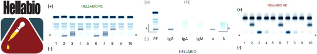
Urine analysis is an essential component in the investigation of patients with paraproteinaemia or with suspected B cell malignancies. An important feature used to help distinguishing malignant from non-malignant conditions is the finding of immunoglobulin fragments produced by tumour cells. The fragments, which may not be detectable on serum electrophoresis separations, are usually of lower molecular weight than intact immunoglobulin molecules. These fragments pass readily through the kidney, and may be clearly visible in the urine due to the concentration effect. The finding of Bence-Jones proteins (BJP) therefore provides a high index of suspicion for malignancy, although it does occur in apparently benign conditions. Even low concentrations (10mg/l) may be significant and therefore concentrated urine (at least 100-fold concentration of early-morning urine is preferable) must be run in the serum systems. Whatever system is used, a trace of albumin must be visible in all urine samples; if this is not the case, the sample should be re-run or further concentrated.
Where a BJP is present in significant concentrations (>100mg/l) with no accompanying glomerular or tubular proteinuria, detection is straightforward and the immunofixation identification step is unequivocal. However, the renal damage associated with BJ proteinuria, frequently results in complex, non-standard patterns requiring immunofixation to resolve the possible presence of BJP.
A low concentration of BJP may also accompany significant glomerular proteinuria in patients with light chain renal amyloidosis; the urine of any such patient should be investigated by immunofixation even in the absence of a band suggestive of BJP. Patients with serum paraproteins may show a "leak" of the serum paraprotein into the urine. This may occur with or without Bence Jones protein and immunofixation is essential to distinguish these.
A number of other proteins may appear as discrete bands on urine electrophoretic separations, particularly where there is an element of tubular proteinuria. These include the α- and β-microglobulins, lysozyme (migrating in the slow gamma region), degraded fragments of glomerular origin and rarely seminal fluid proteins. In some samples the β2-microglobulin will be present in high concentrations and give a very prominent band.
Normal Values
- Protein is not normally found in large quantities in the urine.
Urine protein is roughly divided into urine albumin (less than 50 mg/dL) and globulins. Urine protein electrophoresis may be recommended to help determine the cause of protein in the urine or as a screening test to measure the various proteins in urine.
What abnormal results mean
- Kidney failure
- Decreased kidney function
- Diabetic nephropathy
- Acute inflammation
- Nephrotic syndrome
- Acute urinary tract infection
- Multiple myeloma
- Amyloidosis
Special considerations: Drugs that can affect the measurement of proteins include chlorpromazine, corticosteroids, isoniazid, neomycin, phenacemide, salicylates, sulfonamides, and tolbutamide. Never discontinue any medication without consulting your health care provider.
Drugs that can increase measurements include acetazolamide, aminoglycosides, amphotericin B, cephalosporins, colistin, griseofulvin, lithium, methicillin, nafcillin, nephrotoxic drugs (such as arsenicals, gold salts), oxacillin, penicillamine, penicillin G, phenazopyridine, polymyxin B, salicylates, sulfonamides, tolbutamide, and viomycin.

 Login
Login Forgotten password